COTW 3/7/22: 24 year old female with abdominal pain
A healthy non-pregnant 24 year old female presented to the emergency department with 2 days of lower abdominal pain. Pain was constant, non-radiating and associated with nausea and non-bilious non-bloody emesis. Vital signs: normal. Blood work revealed leukocytosis. Urine pregnancy test negative. Physical exam revealed a comfortable appearing patient with tenderness to palpation along right lower quadrant; non-acute abdomen. CT abdomen and pelvis radiology report: unremarkable, appendix not seen.
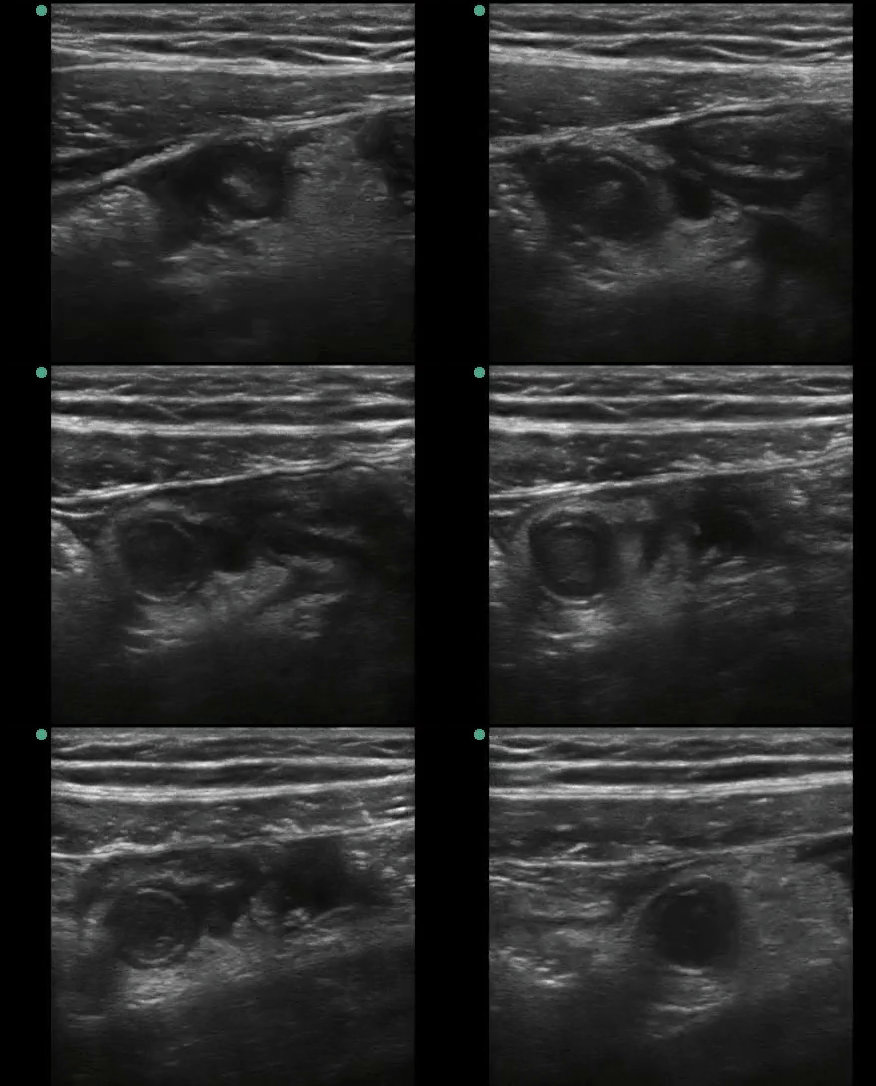
Point of care transvaginal ultrasound showed the following:
A tubular structure was visualized in the right lower quadrant. Cross sectional diameter measured 11.5 mm.
Structure was non-peristalsing, non-compressible and was hyperemic vascular flow as shown above
Surgical evaluation was pursued given concerns for acute appendicitis. Given lack of CT findings of appendicitis, a decision was made to withhold surgical intervention and patient was discharged home with return instructions. Patient returned the following day with worsening pain. CT was repeated which showed acute appendicitis. Patient had an eventful recovery.
Discussion
Acute abdomen in non-pregnant females is a challenging diagnosis
Gynecological and non-gynecological emergencies share similar clinical manifestations
Physical exams are notoriously unreliable
Point-of-care ultrasound allows rapid, safe and accurate evaluation. It adds diagnostic value when evaluating acute abdomen in young female patients
Although CT carries a higher diagnostic accuracy for acute appendicitis compared to ultrasound, it can be limited in case of a low-lying appendix within the pelvic cavity
Background
Most common surgical abdominal emergency worlwide
Diagnosis may be challenging due to atypical presentations
Mimics other pathologies
Enterocolitis, Crohn’s, diverticulitis, omental infarcts, mesenteric adenitis, pyelonephritis, ureterolithiasis, ovarian torsion, ectopic pregnancy, PID, hemorrhagic ovarian cyst, among others
Appendicitis & POCUS
Sensitivity 86%, Specificity 91%
Technique:
In pediatrics and thin patients, use the high-frequency (aka. linear) probe
In adults with larger habitus, use the curvilinear probe
How to find it!
Place the patient in supine position
Place probe at the point of maximum tenderness
If you can’t find it…
Move laterally to identify the ascending colon and lateral abdominal wall
Move the transducer on the lateral border of the cecum
most lateral structure in the RLQ
gas filled (think dirty shadows!)
identify the haustra on the ascending colon caudally
Move transducer medially, across the psoas muscle and iliac vessels (these are your landmarks!)
With the psoas and iliacs kept in view, slide the transducer up (towards the umbilicus) and down (towards the pelvis)
Troubleshooting
Place patient supine
Place patient in left posterior oblique position while applying pressure dorsally on patient’s RLQ from the back
Technique
Use graded compression until landmarks are visualized
It is found in-between or anterior to the psoas/iliac vessels
Normal appendix: blind-ended tubular structure
Sonographic findings in acute appendicitis
Primary:
Non-compressible blind-ended tubular structure
might be compressible if it’s perforated
Lacks peristalsis
Outer diameter measures > 6mm
Wall diameter > 3mm
Secondary:
Appendicolith/fecalith within the lumen
Free fluid surrounding the appendix
Ring of fire = increased vascular flow using color doppler
Bowel wall edema
Diameter 11.1 mm
Periappendiceal edema
Secondary findings for appendicitis: free fluid surrounding blind-ended tubular structure
Key Points
Evaluating lower abdominal pain in young female patients continues to be challenging for emergency physicians
Transvaginal ultrasound adds diagnositic value on low-lying appendices
Optimize technique using compression approach, placing patient in left posterior oblique position
Remember a perforated appendix could be compressible


