COTW 2/28/22: 45 year old male with shortness of breath
A 45 year old male with history of CVA, CHF (EF= 50% on last ECHO done 6 months prior), diabetes, hypertension and hyperlipidemia presents to the ED with 1 week of shortness of breath. Patient denied chest pain, palpitations, fever, cough and diaphoresis. Patient reported medication as well as diet restriction non-compliance and a 15 lbs weight gain.
Vital signs: BP 145/65, HR 75, RR 19, SpO2 94% RA.
Physical exam: Patient is no distress, speaking in full sentences. No JVD noted. Lungs clear to auscultation. Lower extremity +2 pitting edema noted. Blood work was remarkable for a BNP of 500 but close to patient’s baseline. Patient was given 40mg of IV lasix in the ED. Patient was deemed hemodynamically stable. His work up was reported as “unremarkable except elevated BNP”. ED physician’s plan was to discharge patient home with close PCP and cardiology follow up. Before discharge, ultrasound fellow performed a bedside ECHO. . .
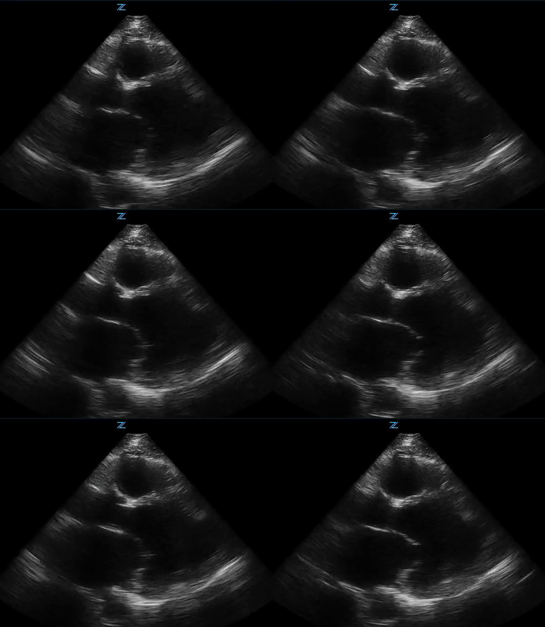
Point of care ultrasound revealed the following:
Long axis view (PLAX) shows global hypokinesis and poor ejection fraction. Visual estimate of EF was 10%.
Apical 4-chamber view shows a large apical thrombus!
Patient also had evidence of pulmonary edema on lung ultrasound and IVC was plethoric with minimal variation with respiration.
Heart Failure
Heart failure is a common illness that presents to the Emergency Department. It affects approximately 2% of the general population in the United States and it encompasses 20% of all hospitalizations.
Precipitants of acute decompensated heart failure (ADHF):
Acute coronary syndrome
Pulmonary Embolism
Valvular disorder: severe aortic stenosis, acute MR, mitral stenosis, chordae rupture
Aortic dissection
Arrhythmias: most common is A-fib
Infection: endo/myo/pericarditis, PNA, other
Anemia
Uncontrolled HTN
COPD
Medication/diet non-compliance
Alcohol/illicit drug use
Endocrine pathology
Uncontrolled diabetes
Thyroid
Medications: NSAID, CCB/BB toxicity
Common symptoms:
Dyspnea on exertion: most sensitive
Orthopnea
Exercise intolerance
Physical exam:
Wheezing
Rales
JVD
S3
Lower extremity edema
Tachypnea, hypoxemia
Work up and what to look for:
EKG: ischemia, dysrhythmias, heart strain pattern (pulmonary embolism, cor pulmonale)
CBC: anemia
CMP: renal function, electrolytes (including Ca/Mag/Phos)
Trop, BNP
CXR: 15-20% of patients with ADHF will lack pulmonary congestion on CXR. Findings may lag 12 hours from onset of symptoms. Sensitivity 60%. Patients with right heart failure might have a normal size heart.
Other findings: cardiomegaly, pleural effusions, Kerley B lines, peribronchial cuffing, perihilar congestion, cephalization
The Approach
Point of care ultrasound is an invaluable tool when evaluating patients with suspicion of ADHF. With POCUS, one can obtain qualitative measurement of patient’s heart function . Evaluation of acute decompensated heart failure include: ECHO, lungs (b-lines) and IVC.
** Remember pulmonary edema requires presence of 3 or more B-lines in 2 or more fields bilaterally **
Image Acquisition
At least 2 views!
PLAX: global function
PSAX: regional wall motion abnormalities, right heart strain
Apical: global function, apex, right heart strain, valves
SubX: global function, pericardium
Management
Treat underlying precipitant
Non-hypotensive patients: NIPPV, nitroglycerin, diuretics (after NIPPV and nitro!)
Hypotensive patients: inotropic support (dobutamine, milrinone), vasopressors (norepinephrine, epinephrine, vasopressin)
Back to our case
Performing bedside ultrasound provided information that drastically changed disposition. Not only patient was found with a large apical thrombus, his ejection fraction was significantly diminished when compared to his previous ECHO. Patient was admitted for medical optimization which included systemic anticoagulation, increase in diuretic dose, strict I&O and diet modification. TTE was performed by cardiology team which confirmed large apical thrombus. EF was quantified as 5-10% with severe global hypokinesis. Patient had an uneventful hospitalization and was discharged home few days later.
Teaching Points!
Approach to patients with concerns of ADHF: ECHO, lungs, IVC
CXR could be normal in early ADHF
POCUS increases diagnostic accuracy and influences physician decision making

