COTW 3/12/23: 71M with chest pain after a fall
A 71M presents to the ED with R sided lateral and anterior chest wall pain following a mechanical fall yesterday. Pain is worse with movement, palpation and deep inspiration. He has been using Tylenol, without significant improvement but he is unable to use NSAIDs because of a prior ulcer. He is otherwise fairly healthy and independent, however for the last day his pain has prevented him from getting out of bed.
The triage nurse orders basic labs, EKG and 2 view chest X-ray when he arrives, however it is after 7P so you’re responsible for interpreting the image. It’s midnight when you open the xray below.
Interpretation? Admittedly my radiology reading skills are mediocre at best, but rib fractures can be extremely subtle, particularly when the occur below the diaphragm. Studies show that US is more sensitive and specific than xrays in detecting rib fractures from blunt trauma; X-rays may miss up to 50% of rib fractures even on oblique views. (Side note, I didn’t learn about oblique chest views ie “rib xrays” until my last week of residency. Often patients with rib pain will have a standard 1 or 2V XR already completed by the time you see them but if you are worried about rib fractures, particularly inferior or lateral ribs, the oblique views can help a lot).
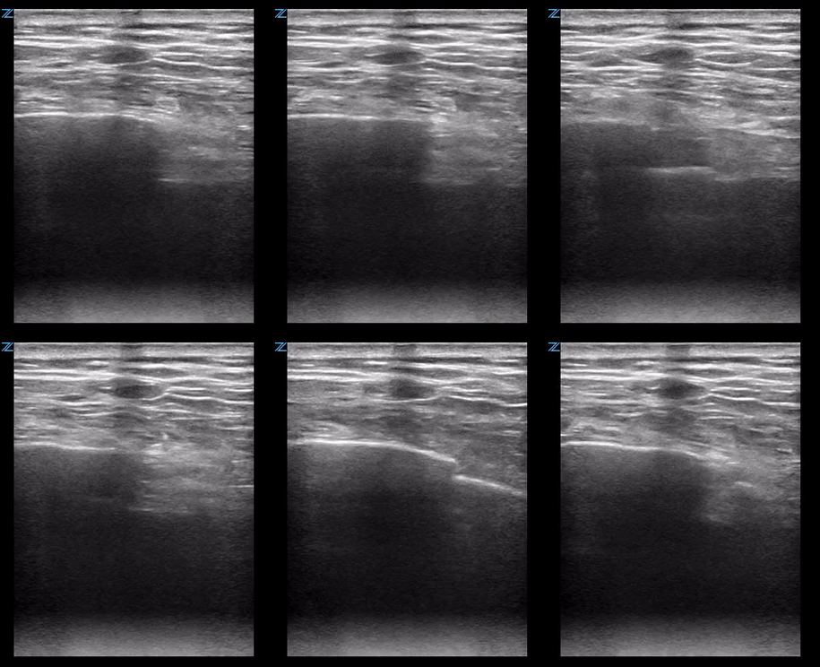
After a few minutes of squinting at the xray above, you realize it would take me a lot less time to simply ultrasound this patient’s ribs than it’s taking to interpret the image. So you get take the US with you as you go in for your initial evaluation and you get the images below.
Rib ultrasound is fairly simple. Ask where the patient is having the most pain, place the linear probe on top of and parallel to the rib(s) in that area. Identify the hyperechoic cortex and the rib shadowing below, the follow the cortex along the longitudinal path of the rib looking for distributions in the cortex.
The key is to remember that the ribs are obliquely oriented so when placing your probe on the patient you will need to angle it so it stays parallel to the path of the ribs, otherwise you may only catch slices of the bone and not see the full line of the cortex as above. If you’re having trouble identifying the ribs particularly in patients with a large amount of soft tissue, it can be helpful to place the probe sagittally and mark where you see each rib then reorient transversely to follow each rib longitudinally.
ULTRASOUND TO DETECT RIB FRACTURES:
CONS
May miss rib fractures, especially if not doing a complete thoracic scan or if patient can’t tell you where their pain is
May be more painful for the patient
User dependent
May take more time than an xray if you’re doing a full exam
PROS
Improved sensitive and specificity compared to xray
Faster, especially if concentrating on the area(s) of maximal tenderness
May be more accessible in low resource settings
Less radiation (important for kids) and less cost
Allows quick, simultaneous assessment for underlying lung pathology.
By now you’re asking, why does this matter? Will missing a subtle rib fracture change management? The answer is probably not, but it may give you and more importantly the patient some peace of mind knowing the definitive answer.
Now, the most important question, what are you going to do about your patient’s pain?
The answer: SERRATUS ANTERIOR PLANE BLOCK
This is one of those nerve blocks that is super simple but can actually make a big difference in disposition and outcome (think frail old person, high fall risk and mildly demented at baseline even without narcotics, also high risk of getting pneumonia).
ED Indications: mostly pain from rib fractures, thoracostomy tube placement, thoracic lacerations or pain from thoracic blunt trauma
Equipment:
15-20cc anesthetic (usually bupivicaine 0.5%, ok to use lidocaine if you need if just doing this for tube insertion or lac repair, although using bupiv can help with the post procedural soreness).
10-15cc normal saline to dilute the anesthetic
20-22g (spinal) needle
ultrasound, sterile cleaning solution
Anatomy:
Your target is the lateral cutaneous branch of the thoracic intercostal nerve which runs between the latissimus dorsi and the serratus anterior muscles. Note, the thoracodorsal artery also runs in this plane so make sure you use color doppler to confirm this artery is not in the trajectory of your needle prior to the procedure.
https://www.acepnow.com/article/ultrasound-guided-serratus-anterior-plane-block-can-help-avoid-opioid-use-patients-rib-fractures/
Procedure:
1) Place the patient in the lateral decubitus position with the affected side up. If possible have them abduct their arm upwards over their head or give themselves a hug with their arm adducted across their chest to move it out of the way. Place the probe transversely in the mid axillary region with probe maker facing forward.
https://www.acepnow.com/article/ultrasound-guided-serratus-anterior-plane-block-can-help-avoid-opioid-use-patients-rib-fractures/
http://highlandultrasound.com/serratus-anterior-plane-easy
2) Position the probe until you identify the tail end of the latissimus dorsi (posterior) as it tapers anteriorly into a fascial plane above the serratus anterior. You should see the rib laying below the serratus as well as the pleura between the ribs.
3) Once you’ve identified your landmarks, advance your needle in plane until you penetrate the fascia between the muscles. Insert a small amount of the dilute anesthetic solution to confirm you’re in the correct place then watch the two muscles separate as your inject the rest of the solution.
http://highlandultrasound.com/serratus-anterior-plane-easy
The relative thickness of the the latissimus compared to the serratus will vary based on how far anterior vs posterior you are. The images above were obtained with the probe farther anterior on the patient, however the latissmus becomes thicker and the serratus thinner as you slide the probe marker more towards the posterior axillary line. Since this is a plane block, your injection site can be anywhere along the plane between the two muscles. Remember for pain that is more lateral you will likely need to start farther backwards, however in general try to find the location where you can see the fascia the best. In the example to the right, you see a thick latissimus above a thin serratus muscle. Another tip is to aim your needle for an injection site above a rib using the rib as a backstop to further ensure you don’t penetrate the pleura.
Congratulations! Another day successfully diagnosing and treating your patient’s condition with just your ultrasound.


