COTW 3/28 79 year old with abdominal pain
A 79 year old female with a history of several prior abdominal surgeries presents with abdominal pain and vomiting. She appears uncomfortable on exam and you already know that she has a trip to the donut of truth in her future.
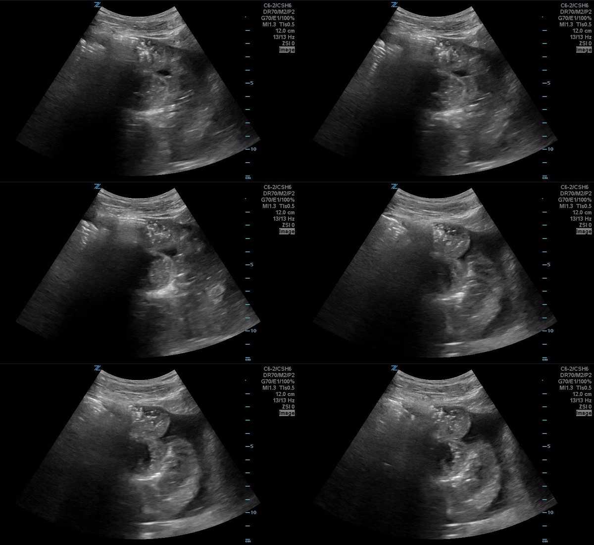
While awaiting the truth-seers input, you decide to place an ultrasound on her belly and see this.
Small bowel and free fluid
The above represents a small bowel obstruction. In the vast majority of cases we really shouldn’t be able to see bowel and if you do see bowel, you should be suspicious that some sort of pathology might be present. This is due to the fact that bowel is largely gas filled and gas doesn’t transmit sound well.
In this case, we see a couple of features of small bowel obstruction. The first is that the bowel wall diameter is greater than 2.5 cm. Even without doing a formal measurement we can see that is the case. There is also clear “to and fro” peristalsis where the feculent material inside the obstruction tries to peristalse forward, but then gets propelled backwards because of the obstruction. Below is a little clearer example of that.
To and Fro peristalsis
In the above image you can also see the “keyboard sign” which are the plicae circularis being highlighted by all of the fluid within the bowel. In comparison, a large bowel obstruction will instead have echogenic arcs of bowel giving it a more segmental appearence.
Large Bowel Obstruction
This scan is very easy to perform, start in the RLQ and just lawn mower up and down the abdomen with the curvilinear probe. It won’t replace cross sectional imaging, but identifying bowel obstruction on pocus can provide you early diagnostic insight.


