COTW 9/8/22: A 74yo F with R arm pain and swelling
A 74yo F presents to the ED with R arm pain and swelling for 2 days. Had a pacemaker placed on the R side about 9 days ago. Pacemaker placement was complicated by ventricular perforation and pericardial effusion which was resolved with pericardial drain placement. Sent home, told to hold her blood thinners for 2 weeks.
Ultrasound
〰️
Ultrasound 〰️


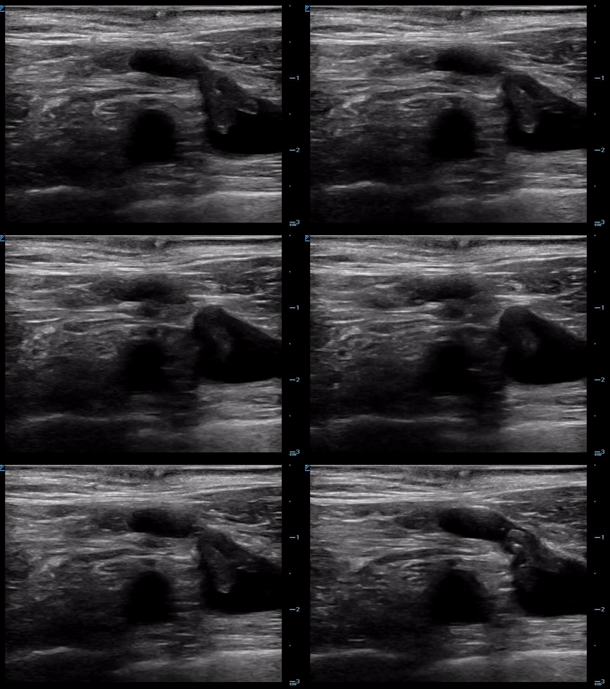
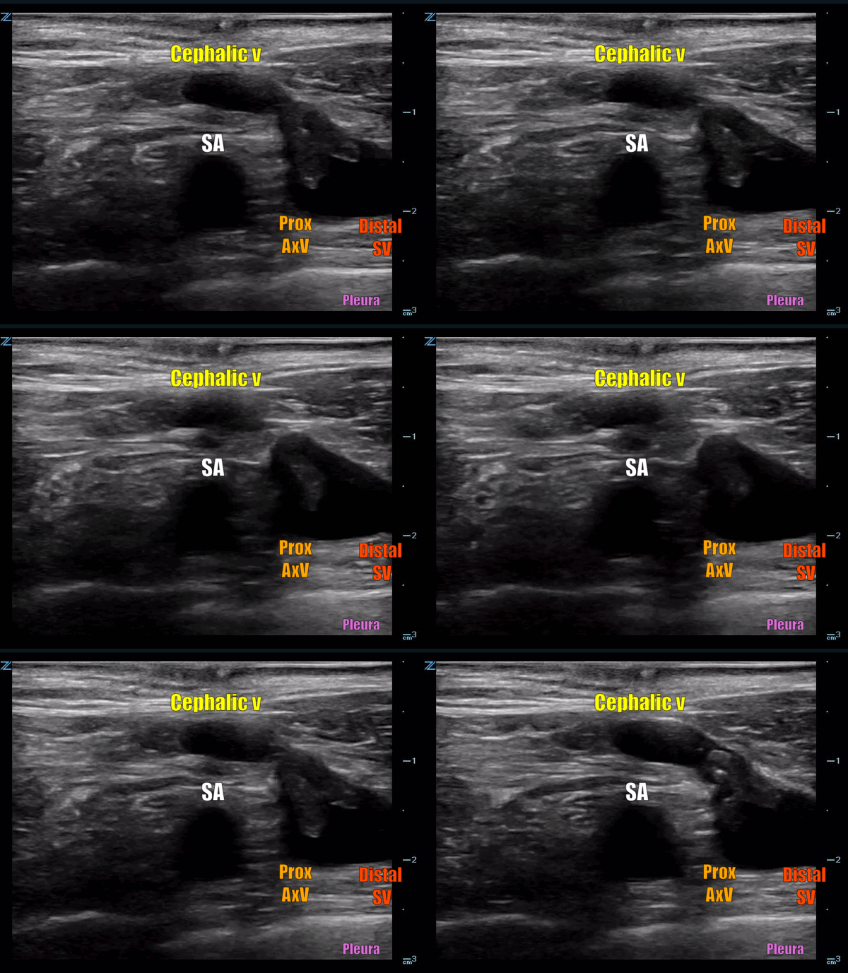
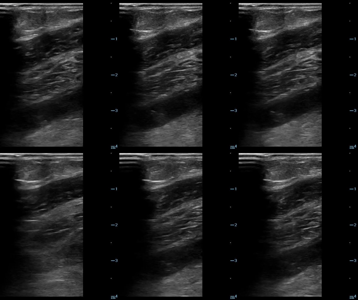
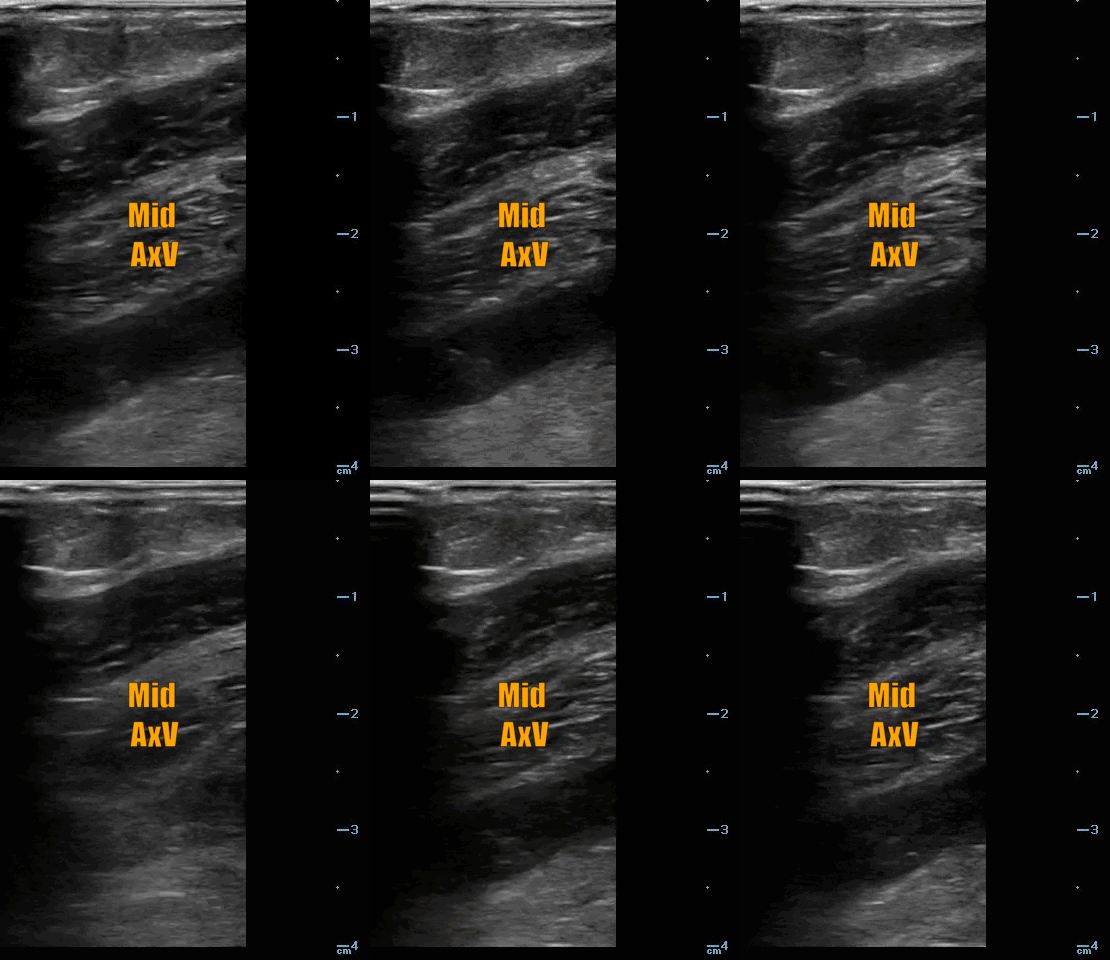
The image on the left above shows normal anatomy as you follow the upper extremity veins down from the IJ to the subclavian vein.
The image on the right shows normal, biphasic doppler flow (A) and abnormal continuous doppler flow (B) through the subclavian vein. Continuous flow is seen with obstruction (thombus).
REFERENCES:
D.S. Giraldo Gutiérrez, J. Bautista Sánchez, R.D. Reyes Patiño. Supraclavicular approach for subclavian vein catheterization in pediatric anesthesia: The reborn of an ancient technique with the ultrasound's assistance, Revista Española de Anestesiología y Reanimación (English Edition), Volume 66, Issue 5, 2019,Pages 267-276, https://doi.org/10.1016/j.redare.2019.01.001
Czihal, Michael & Hoffmann, Ulrich. (2011). Upper extremity deep venous thrombosis. Vascular medicine (London, England). 16. 191-202. 10.1177/1358863X10395657.
Miller A, Vermeulen M. Ultrasound-Guided Subclavian Vein Cannulation, the Vessel to Remember. EMResident. 4/9/2019. https://www.acep.org/sonoguide/basic/dvt/


