
COTW 11/6/22 62F presenting for shortness of breath
62F no known past medical history presents with progressive dyspnea on exertion now with shortness of breath at rest.
BP: 88/53 (MAP 64), RR 24 SpO2 92% on RA,
Patient is fatigued appearing, mildly tachypneic with poor inspiratory effort but no significant respiratory distress. Obese abdomen, protuberant but no tenderness. 1+ pitting edema bilaterally. Denies lightheadedness or dizziness, mental status is normal.
You start the patient on 2L NC with improvement in her oxygenation. While your nurse is getting an IV, you complete a RUSH exam to help figure out why the patient is hypotensive. Your cardiac views shos grossly normal EF, no pericardial effusion, collapsible IVC. RUQ view below:

DIAGNOSIS?

HEPATIC HYDROTHORAX:
Pleural effusion (usually >500mL) in patient with cirrhosis without any other underlying pathology that could cause an effusion (ie no cardiac, pulmonary or pleural disease)
Occurs in 5-15% of patients with cirrhosis
Ascites fluid is sucked through small diaphragmatic holes into the pleural space largely driven by negative intrathoracic pressure (therefore some patients with hepatic hydrothorax don’t actually have ascites)
Right sided hepatic hydrothorax thought to be more common (73-85% of patients) because the left hemidiaphragm is thicker and more muscular. Isolated left sided hydrothorax occurs in about 13-17% of patients, whereas 8-24% have bilateral effusions.
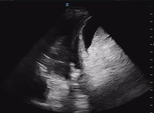
A second example of hepatic hydrothorax, again with fluid above and below the diaphragm in the context of a nodular appearing liver.
What are the ultrasound characteristics of a cirrhotic liver?
Cirrhosis is characterized by hepatocellular necrosis leading to fibrosis, nodular regeneration and distortion of hepatic architecture which can be identified on ultrasound by:
nodular or bumpy appearing liver edge
hypoechoic nodules seen within the parenchyma
overall coarse, heterogenous appearance
may have signs of portal hypertension (distended portal vein)
may also have ascites and splenomegaly
As a very efficient physician, you have the time to sneak in a quick diagnostic thoracentesis. Are you expecting a transudative or exudative effusion, and what are the criteria to distinguish?

Your labs confirm your diagnosis. Given the collapsible IVC, you give your patient small fluid boluses with improvement in her hypotension. Medicine gladly accepts your admission.