COTW 10/9/22 55 yo male presenting with shortness of breath
55 year old male history of CHF, COPD, DM presents for evaluation of shortness of breath. He states that over the past week he has had worsening shortness of breath, subjective fevers, orthopnea, slightly more swollen legs. His vitals are BP 147/82 HR 90 SpO2 86 RR 20 T 99.4. His pulmonary exam is notable for apical wheezes and basilar crackles. The rest of his exam is notable for trace pedal edema. To better evaluate his dyspnea you decide to perform lung ultrasound.
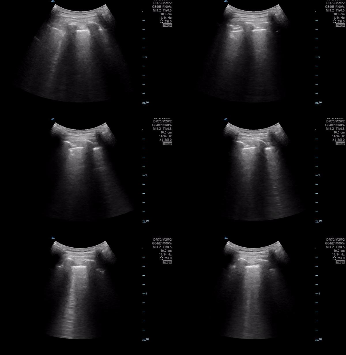
Scattered B lines-appreciated bilaterally
You notice the above pattern of B lines across the anterior precordium bilaterally. However at the lung bases you see the following.
While more prominent on the left side, above is what is called a “shred sign”. You can see above that the bright white pleura that is normally smooth looks like it has been crinkled like tissue paper. What you are seeing are sub-pleural consolidations and their irregular borders with more aerated lungs. This is suggestive of a pneumonia. This case also highlights how if you aren’t systematic with lung ultrasound, you will miss things!!!
Ideally, for proper lung ultrasound you want to get three views(see below). Getting all three of these views will ensure that you do not miss pathology.
Case Resolution: After performing lung ultrasound you decided to order antibiotics. His chest x ray is relatively unremarkable however his d-dimer was elevated so a CTPE was ordered. No PE was seen but bilateral pneumonias were visualized.


