
COTW 4/12/20: A 13 y/o M with RLQ abdominal pain
A previously healthy 13 y/o M is brought for few days of vague abdominal pain that has now concentrated over RLQ of abdomen. Symptoms are associated with fever, nausea, and vomiting… so you put a probe on the belly:

At the RQL, many lymph nodes may be seen. No clear sign of appy here, but the suspicion is quite high…
As we scanned superiorly, there was still considerable tenderness as we arrived to the RUQ, so we kept looking…

Found something! This fluid filled structure is located well into the RUQ. In fact, that’s the liver just to the left on the screen! As evidenced on this clip, it is not compressible by pressure.

Short axis measurement well over the upper limit of normal for an appendix of 6mm.
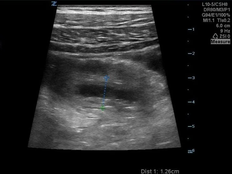
Longitudinal view shows similar measurements with surrounding edema.
So why couldn’t we find it in the RLQ?
Here’s a clip showing how it dives deep behind the ascending colon, where it becomes obscured by intraluminal air, making it difficult to visualize it in the usual location.

Fortunately, these images clinched the diagnosis and the patient was taken to the OR for a successful, uncomplicated laparoscopic appendectomy, without the need for ionizing radiation from CT scanning.
So just remember, that although we typically look in the RLQ, the location of the tip of the appendix is highly variable as it can be located retrocecal, pelvic, pre- or post-ileal, paracecal, or subcecal.
So if your suspicion is high, be aggressive in looking for it with ultrasound in the right population.