COTW September 4th: A 90 year old man with chest pain.
This is a case of a 90 year old male with abrupt onset chest pain radiating to his back. He was mildly hypertensive and with a normal EKG.
Bedside echo as follows:
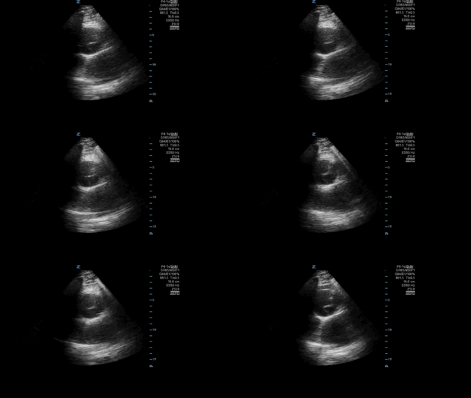
Parasternal long axis. Notice the dilated aortic root and intimal flap. This aortic root measured 4.98 cm.
This is a high parasternal short axis view. You can also see the intimal flap here.
Patient’s blood pressure was managed acutely, sent to CT scanner, Thoracic surgery was consulted immediately.
Learning points:
-Aortic root size >4 concerning for aortic root aneurysm and/or dissection
-Always evaluate for pericardial effusion when concerned for dissection or if there is evidence of inferior/right sided STEMI on EKG
-Utilize all your views (parasternal long axis, high parasternal short axis, subxiphoid). Suprasternal notch view is also a great way to pick up aortic dissection on POCUS.

